Parts of the microscope review
As a doctor is conversant with the use of a stethoscope, the laboratory scientist is to the microscope. For a microscope to function effectively, each and every part must be in good working good condition to enable you to magnify and view your specimen. When one part of the microscope is in good working condition and another part is faulty, the microscope becomes a white elephant. Parts of the microscope review will enable you to teach students who do not understand the basic concept of the microscope and also help colleagues understand the basic principle of the microscope. Take a look at the attune flow cytometer.
The function of the parts of the microscope
Using the microscope as a beginner will pose a challenge to you. This blog is here to reduce or eliminate the challenge since it will explain the various function of the parts of the microscope.
Lens
A lens is simply a glass material that is used to magnify small objects. The same principle is used in a microscope. It magnifies the specimen to be viewed. The type of lens used in the microscope is convex. The microscope is made of two lenses. The eyepiece lens is also known as the ocular lens and the objective lens.
Objective lens
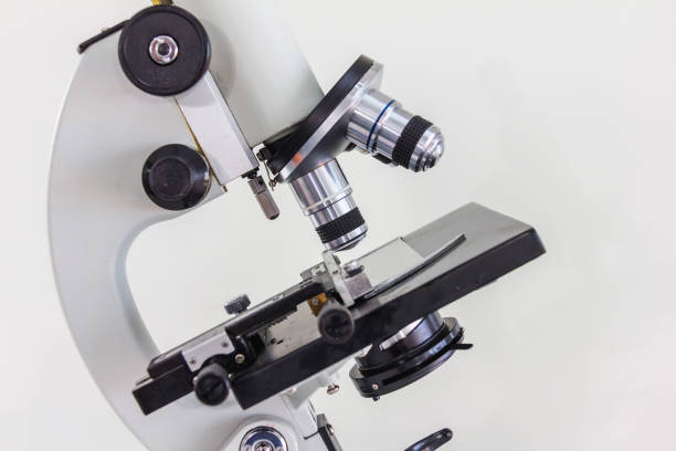
The objective lens is the first magnifying lens. It magnifies the specimen. It consists of four lenses though some may contain three. The lenses are a 4x-scanning lens, 10x-low power lens, 40x-high power lens, and 100x-oil immersion lens. They are hooked to the revolving nosepiece.
Ocular lens
It is popularly known as the eyepiece lens. It does not only serve as a way for viewing the specimen but also as another magnifying lens. It further magnifies the object that has already been magnified by the ocular lens giving the image or clear view. The range of magnification of the eyepiece ranges from 5x to 30x with most being 10x.
The magnification of the microscope is the product of the objective lens and ocular lens. For example, a specimen viewed under a 100x objective lens and 10x ocular lens will have a magnification of x1000.
Diopter adjustment
It is used to alter the focus of one ocular lens to give the right vision for both eyes.
Focus knobs
These are the parts of the microscope that make one a great microscopist. These are the coarse focus and fine focus
Coarse adjustment knob
It is used for moving the stage up and down. This allows the specimen to be brought close to the objective lens to allow perfect magnification.
Fine adjustment knob
As the name indicates, is used to make the image fine and clear. After the stage has been raised and brought close to the ocular lens, the fine adjustment knob is turned to see the detailed image magnified by both the objective and ocular lens.
The fine adjustment knob is mostly smaller than the coarse adjustment knob.
Light source
This is simply the source of light of the microscope to enable viewing of the specimen. It used to be a mirror that directs an external source of light to the objective lens. Most microscopes now use the light microscope with incandescent lamps. Modern microscopes like Zeiss can even control the intensity of the source of light.
Condenser
The condenser is used to direct and concentrate light onto the sample, As the light is coming from the source, It may spread to parts of the slide which have no sample or specimen. The condenser makes it easy by directing the light to help you view your sample.
Diaphragm/ Iris
This is a small lever that controls the amount of light that comes out of the condenser. Pushing the lever left to right can increase or decrease the light escaping from the condenser.
Stage
This is the platform where the slide is placed for viewing. Attached to the stage are the stage clips for holding slides firmly to the stage to prevent falling when moving the stage. Take a look at the lazle blood pressure monitor.
Stage control knobs
The stage control knobs move the stage forward, backward, left, and right. Sometimes the stage may be away from the light so the knob comes in to bring the sample right above the light. Again, one cannot diagnose a disease by viewing only part of the field. You have to view different fields and this can be achieved by using the stage control knobs the navigate the field to find and diagnose the suspected disease.
Aperture
This is the small hole at the center of the stage that the light passes through to reach the specimen.
Switch
This is located at the base of the microscope to turn the microscope on and off.
Revolving nosepiece
This is what holds the objective lens in position. It can be rotated to select the right lens needed to execute the job.
Body tube
It connects the ocular lens to the objective lens.
Arm
It joins the base of the microscope to the body tube.
Base
It is the platform on which the microscope is built. It provides support. It houses [arts of the microscope like the light source and switch.
Parts of the microscope review- Questions
1. The lenses of the microscope are
a. ocular lens and objective lens b, diaphragm and ocular lens c. objective lens and tris
2. What is the other name of the 4x objective lens?
a. scanning lens b. high power lens c. low power lens d. oil immersion lens
3. What is the other name of the 10x objective lens?
a. scanning lens b. high power lens c. low power lens d. oil immersion lens
4. What is the other name of the 40x objective lens?
a. high power lens b. oil immersion lens c. scanning lens
5. Which part of the microscope controls the upward and downward movement of the stage?
a. stage control knobs b. fine adjustment knob c. diaphragm d. coarse adjustment knob
6. ………………… gives a detailed image of the specimen.
a. coarse adjustment knob b. stage control knob c. fine control knob d. condenser
7. What is the magnification of a sample viewed under an objective lens of 40x and an ocular lens of 10x?
a. 4000x b.400x c. 40.0x d. 4.0x
8. …………………… moves the stage from right to left.
a. stage control knob b. fine adjustment knob c. diaphragm, d. coarse adjustment knob
9. The …………….. controls the amount of light before it reaches the specimen
a, light source b. ocular lens c. diaphragm d. condenser
10. The ………… directs and concentrates light onto the specimen
a. light source b. diaphragm c. ocular lens d. condenser
Answers to parts of the microscope review
- a
- a
- c
- a
- d
- c
- b
- a
- c
- d
Conclusion
This blog on parts of the microscope review will not only guide beginners but also students which help them operate the microscope with ease and avoid most challenges beginners face.