With the help of microscopy, we can now see and examine the minute intricacies of cells and tissues, which has completely changed how we think about the microscopic world. In this article, we delve into the fascinating world of inverted fluorescence microscope. We will decipher the underlying mechanisms, go over the elements, and look at the various uses that make this method a mainstay of biological research.
What is an inverted fluorescence microscope?
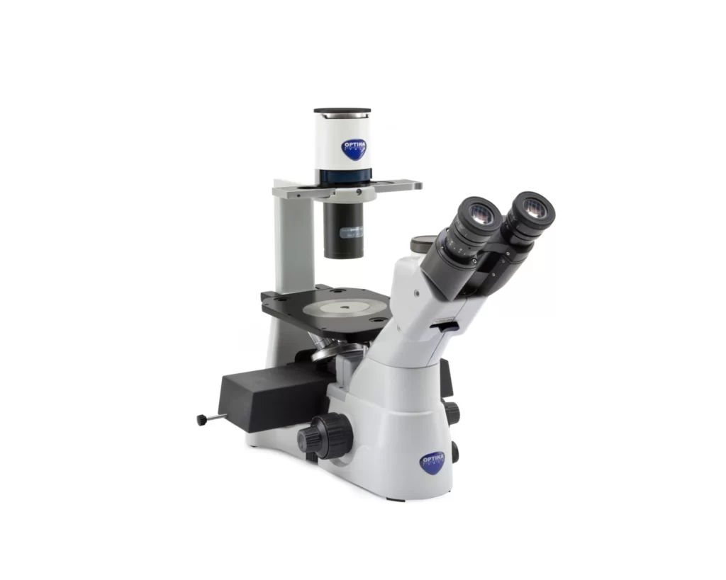
An inverted fluorescence microscope is a unique microscope that differs from the conventional upright design of microscopes with the objective lens placed below the specimen while observing the images from the ocular lens in the eyepiece above. The inverted shape makes it particularly ideal for live cell imaging and other applications involving complex sample settings. It permits the evaluation of samples in or on slides, Petri dishes, multiwell plates, or other containers.
What is the principle of the Inverted Fluorescence Microscope?
The excitation light is produced by the inverted fluorescence microscope at a particular excitation wavelength, focused onto the sample by condenser lenses, and then absorbed by fluorescent molecules (fluorophores). Fluorescence, which provides information about labeled structures or molecules, is released when the excited electrons in fluorophores return to their ground state. The objective lens collects the fluorescence that is released, and fluorescence filter sets are used to separate it from the excitation light. Researchers can examine cellular activities by converting detected fluorescence signals into digital images for real-time observation and analysis. The microscope’s inverted form enables the observation of samples in multiwell plates or culture dishes, making it appropriate for live cell imaging and complex settings. Do not forget to read about the attune flow cytometer.
Components of the Inverted Fluorescence Microscope
- Base and stand- The base provides support for the microscope while the stand holds the other parts of the microscope.
- Light source- It provides light for the excitation of molecules and it enables the viewing of specimens.
- Condenser- It directs the light unto the specimen.
- Objective lens- It is found below the stage and magnifies the image and allows emitted fluorescence to pass through
- Excitation filter- It provides the excitation light at the precise wavelength needed to stimulate the fluorophores in the sample.
- Emission filter: It selectively transmits the fluorescence that has been produced while blocking the excitation light and other undesirable wavelengths.
- Sample stage- This is the platform where the specimen is placed.
- Eyepiece/ Ocular lens- Some inverted fluorescence microscopes have eyepieces that contain an ocular lens that further magnifies the image magnified by the objective lens.
- Coarse adjustment knob- It controls the upward and downward movement of the stage.
- Fine adjustment knob- It is used to focus the image to appear sharp and clearer.
- Detector– Modern microscopes have cameras to capture digital images or use the aid of a specialized photodetector.
Applications of the Inverted Fluorescent Microscope
- Live Cell Imaging: Samples in culture dishes, multiwell plates, or other containers can be examined using the microscope’s inverted architecture. As a result, it is especially well suited for live cell imaging since it allows for the continuous observation of cells in their natural setting. Additionally, the inverted structure makes it simple to access the material during tests for manipulation or intervention.
- Sample handling flexibility: The inverted microscope can handle a variety of sample sizes and types. It is capable of handling 3D cell cultures, spheroids, and organoids as well as thick samples like tissue sections. It is useful for studying intricate biological systems and cellular processes because of the flexibility in sample handling.
- Compatibility with Specialized Techniques: The inverted fluorescence microscope is capable of being outfitted with a variety of cutting-edge tools and methods, including confocal microscopy, total internal reflection fluorescence (TIRF) microscopy, fluorescence resonance energy transfer (FRET), and fluorescence recovery after photobleaching (FRAP). These methods give researchers the ability to investigate certain cellular relationships, processes, and dynamic events.
- Integration with Imaging Systems: Real-time imagining, image capture, and analysis are all made possible by the seamless integration of inverted fluorescence microscopes with digital imaging systems. Quantitative measurements, picture processing, and data analysis are made possible through the employment of imaging software and cutting-edge camera technologies.
- Reduced Phototoxicity: In order to preserve the life of cells and physiological behavior during live cell imaging, phototoxicity must be kept to a minimum. By keeping the objective lens away from the sample, the inverted fluorescence microscope minimizes phototoxic effects and permits longer examinations without severe harm to cells.
- Access to Sample Bottom: In an inverted microscope, the objective lens is positioned below the sample, allowing for easy access to the sample’s bottom. For applications like intravital imaging, where the microscope can be placed over clear windows or implanted components in living creatures for comprehensive observation, this is especially helpful.
- High-Quality Imaging: Inverted fluorescent microscopes frequently have high numerical aperture (NA) objectives, enabling outstanding resolution and light-gathering abilities. Images produced as a result are of a high caliber with improved clarity and detail. Superior imaging capability is further enhanced by the use of premium optics and exact focusing mechanisms. Take a look at the lazle blood pressure monitor.
What is the Price of the Microscope?
The price of the microscope may differ due to geographical location, platform for purchasing, brand, specifications, model, and many others. The price usually ranges from $3000.00 to $50,000.00.
Conclusion
The inverted fluorescence microscope effective tool that combines the advantages of fluorescence imaging with the versatility to examine living cells and intricate material. It has developed into a essential tool in numerous biological research domains, aiding in our comprehension of cellular connections, functions, and architectures. Do not forget to read about alcedo blood pressure monitor.