The ability to see inside the complex world of cells, tissues, and organisms makes microscopy a vital instrument in the field of scientific inquiry. The manual examination of slides under a microscope is a staple of traditional microscopy, but thanks to technological improvements, a brand-new marvel has appeared the microscope slide scanner. We shall explore the interesting world of the microscope slide scanner in this blog article, as well as its advantages and effects on numerous academic disciplines. Read on sliding microtomes.
What is a Microscope Slide Scanner?
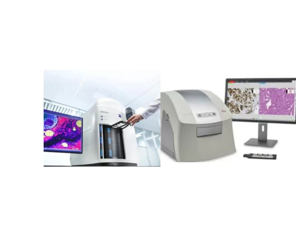
A microscope slide scanner is an innovative tool for automating the digitization of glass microscope slides through cameras, lenses and light sources embedded in the device and displaying them on a computer. It allows researchers and scientists to capture precise and accurate pictures of complete slides showing affected parts of a tissue or cell due to a disease condition using a technology that combines the capabilities of microscopy with advanced imaging technology and computer software.
Other Names
- Slide digitizer
- Slide imaging system
- Slide scanning device
- Virtual slide scanner
- Automated slide scanner
- Digital slide scanner
- Whole slide scanner
- Pathology slide scanner
- Microscopy slide scanner
- Slide imaging workstation
Components of the Microscope Slide Scanner
- Slide Holder: It holds the glass slides firmly on a platform for accurate and high-quality image capture, it makes sure that the alignment and stability are correct.
- Motorized stage– It is an automated platform that places the slide beneath the scanner’s imaging system. It also allows for precise and controlled movement to take numerous pictures at various locations on the slide.
- Digital camera: A top-notch camera or cameras that record slides’ images.
- Lenses: It magnifies the image on the slide.
- Light sources: Just like the traditional microscope, it provides controlled illumination for the slide including LEDs and halogen bulbs.
- Optical route: It consists of mirrors, prisms, and other optical components that allow light to pass through to the slide.
- Image stitching software- It is a specialized software for picture stitching, which automatically combines several photos taken from various fields of view to produce a smooth, high-resolution image of the entire slide
- Computer- It is used to control the operations of the microscope slide scanner and it is connected to the scanner via USB, Ethernet, or other interface for data transfer and communication.
- Cooling system- Advanced microscope slide scanners are embedded with cooling systems to reduce the heat produced as a result of prolonged use of the scanner.
How the Microscope Slide Scanner Works
A microscope slide scanner works on the premise of combining innovative image technology, exact movement control, and complex software algorithms. The basic idea is to take several high-resolution pictures of various fields of view on a microscope slide and combine them to produce a complete and seamless digital depiction of the entire slide. Here is a summary of the fundamental concepts underlying a microscope slide scanner.
The principle of the microscope slide scanner is to digitally capture high-resolution images of slides, a microscope slide scanner makes use of cutting-edge imaging technology, motorized stage movement, and software processing. It uses high-quality optics for accurate imaging and takes numerous pictures as the slide travels methodically. Depending on the capabilities of the scanner, various imaging techniques are employed. Using specialist software, captured photos are edited and stitched together to produce a flawless digital depiction of the complete slide. With this automation, productivity is increased, precise picture reproduction is ensured, remote access to slide data is made possible, and image analysis and collaboration in pathology, research, and teaching are made easier. Take a look at the lazle blood pressure monitor.
Advantages of the Microscope Slide Scanner
- Enhanced Efficiency: By automating the scanning process, microscope slide scanners dramatically increase efficiency. Scientists and pathologists are able to digitize several slides quickly and easily with a slide scanner. Motorized stages accurately move the slide while taking numerous high-resolution pictures and stitching them together. The quicker scanning made possible by this automation leaves more time for data processing, research, and clinical decision-making.
- High-Quality Digital Imaging: Microscope slide scanners create exceptionally high-resolution digital photographs. These scanners are capable of taking clearer, more accurate pictures of minute details because they use cutting-edge cameras, lenses, and illumination systems. Pathologists and researchers may zoom in, inspect complex structures, and spot minor anomalies that could go missed with conventional microscopy thanks to the high-quality images.
- Comprehensive Image Analysis: By utilizing innovative software techniques, the scanners provide strong image analysis tools. These technologies can be used by pathologists and researchers to carry out quantitative measurements, label areas of interest, and use automated analytic methods. This makes it possible to evaluate samples objectively and consistently, improving the precision and repeatability of findings from studies and medical judgments.
- Improved collaboration- Slides that have been digitally preserved can be easily shared across international borders, enabling specialists to work independently. Professionals may efficiently participate in virtual discussions, exchange ideas, and get second opinions by annotating and marking certain areas on the slides. Improved patient care and research outcomes result from this collaborative environment’s promotion of interdisciplinary approaches and acceleration of knowledge sharing. Do not forget to read about the attune flow cytometer.
- Effective data management- Microscope slide scanners deal with the problems posed by slide archiving, retrieval, and storage. Efficient Data Management. Physical storage space and the chance of loss or damage are no longer necessary with digital slides. In order to ensure long-term preservation and convenient retrieval for future use, these digital assets might be housed in secure databases. Furthermore, digitized slides are easily integrated with picture archiving and communication systems (PACS) and laboratory information management systems (LIMS), allowing for effective data administration, retrieval, and searching.
Conclusion
With its multiple advantages and ability to revolutionize numerous academic disciplines, the microscope slide scanner has emerged as an essential tool in scientific research. Slide scanners provide new opportunities for research, teamwork, and data analysis by fusing the power of microscopy with cutting-edge imaging and software capabilities. These tools are poised to transform microscopy in the future and accelerate scientific progress to previously unimaginable levels thanks to their capacity to digitize and preserve priceless samples. Take a look at the toe pulse oximeters.